Patients with heavily calcified coronary artery disease have undergone successful Intravascular Lithotripsy treatment at Cheng Hsin General Hospital
Ptosis surgery can be traced back to the frontalis suspension method published by Dransart in 1880. Although other scholars have proposed different methods, this method is still the first choice for most surgeons when dealing with severe ptosis. However, after surgery, the eyes often cannot be closed completely, and even the problem of drooping eyelids cannot be solved.
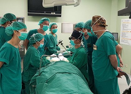
For the treatment of severe eyelid ptosis, it is indeed a difficult challenge to lift the severely drooped eyelid to the normal position, while avoiding the problems caused by the inability to close the eyes after surgery. Based on the anatomical structure of the eye, Cheng Hsin General Hospital uses electromyography to measure the changes of the muscles around the eyes in normal people and patients with ptosis after surgery. The results showed that compared with the normal eyelid without ptosis, the operation of ptosis through the conjunctival incision can avoid destroying the orbicularis oculi muscle and strengthen the function of closing the eyes.
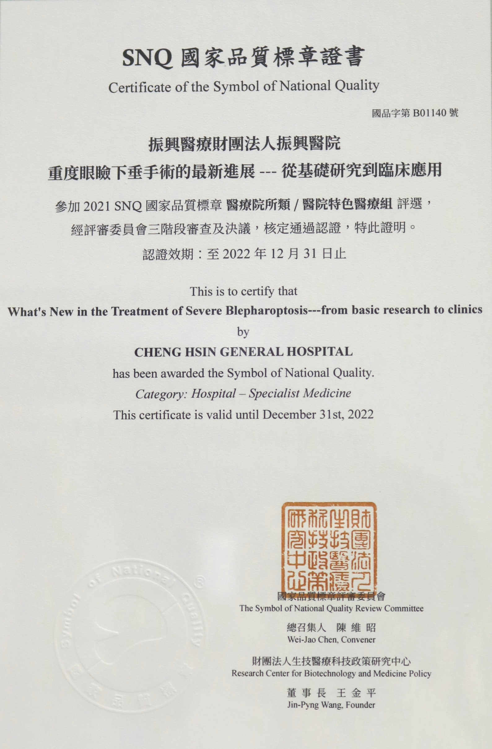
Based on the aforementioned theory, using the incision from the conjunctiva to separate the levator muscle and move it down to the upper edge of the tarsal plate to suture, and to combine the fascia to strengthen the tarsal plate.This surgical method is completely non-marking, not only can treat severe ptosis, but also can recover within a month even if the eyes cannot be fully closed after surgery. This method has been applied to more than 1,215 patients with ptosis, with a patient satisfaction rate of 97.8%. The complete cure rate was 100%, the infection rate and postoperative eyelid insufficiency were all 0%. 17% of the patients were referred for revision by other hospitals. This surgical method has been published in internationally renowned journals of plastic and reconstructive surgery.


 Mon~ Fri 8am~5pm.


